 Loading... Please wait...
Free Shipping on orders over $400
Categories
How to Place ECG Electrodes
Posted on 10/12/2018 10:58:59

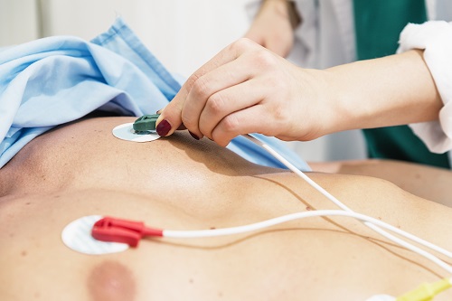
Every time the human heart beats it creates energy in the form of electrical currents. The most common biosensor technology used to collect this data is a painless, non-invasive machine called an electrocardiogram. The ECG machine (also known as EKG machine) measures electrical activity and records it as waveforms. In order to read this data, a medical professional must know how to properly place ECG electrodes onto the patient’s body.
The human body generates electricity every time the heart beats. Using an electrocardiogram machine known as an ECG or EKG, medical professionals are able to measure the health of a patient’s heart. The process for measuring this data is known as the 12-lead ECG test. This test involves the correct placement of 10 ECG electrodes that must be placed in certain places on a patient’s body in order to correctly measure data such heart rate, heart rhythm, and other activity.
The 12-lead ECG test is a vital tool for all sorts of situations, from long-term monitoring in an outpatient care setting to acute-care hospitals and diagnostic centers. Electrocardiograms are often required before undergoing surgery and many adults will have at least one in their lifetime. For the practitioner, it is vital to know to correct ECG placement for each patient.
In order to administer 12 lead ECG placement correctly, it’s important to understand the following:
- How to Prepare for an ECG
- Chest (Precordial) Electrodes and Placement
- Limb Electrodes and Placement
- ECG Electrode Storage, Maintenance and Disposal
- ECG Tips and Best Practices
The ECG uses lead cables and silver chloride electrodes which receive and transmit data when placed firmly on the flat ventral surface of the chest wall. Once the ECG data has been correctly read and interpreted, the reading can help detect and track a multitude of heart conditions like arrhythmias and cardiac ischemia.
The process of measuring and recording this electrical activity over a period of time is called electrocardiography. Patient care centers around the world rely on accurate readings from a specific type of medical technology called the 12-lead electrocardiogram. To properly gather this data, the medical technician must correctly place 10 electrodes on the human body. These 10 electrodes, correctly positioned, provide the 12 perspectives that make the up the 12-lead electrocardiogram.
To clarify any possible confusion, let’s quickly cover the terms ECG and EKG. In the medical literature, you will find that the term electrocardiography is shortened to both ECG and EKG. You might be wondering: ECG vs EKG – what is the difference? As it turns out, they are exactly the same. The difference in the acronyms simply stems from a variation in the German translation of electrocardiography. In this tutorial, we will primarily use the term ECG.
The 12-lead ECG machine is the standard technology used in most medical outpatient and hospital settings, although other variations do exist. Variations of ECG leads include a 3-lead ECG test and a 5-lead ECG test. The 3-Lead ECG uses three electrodes that are typically labeled as white, black, and red, although these colors are not universal because two independent systems for coloring standards exist (for reference, these two systems come to us by way of the International Electrotechnical Commission and the American Heart Association ). The 3-lead ECG machine will track and record the heart’s rhythm but will not reveal sufficient information on heart rate and activity. The 5-lead ECG test uses four limb leads and one precordial lead. While it’s an improvement over the 4-lead ECG test, it is still inferior to the preferred 12-lead ECG.
How to Prepare for an ECG
1.Begin by washing your hands or using hand sanitizer.
2.Explain the purpose for the ECG to the patient as well as what they should expect from the procedure itself. Remind the patient that the procedure is pain-free. Explain that the electrodes will be placed on the skin for just a few moments and overall the testing should last only about 20 minutes.
3.Turn off all non-medical electronic devices, especially smartphones. Interference from these devices can disrupt the ECG readings, making them unreliable.
4.Have the patient lie on a clean, flat and smooth surface in the supine (face up) position. If the patient cannot comfortably lie flat, assist him or her into the semi-fowler position in which the head of the bed or table is elevated 30-45 degrees. Allow the patient to elevate the upper body only if absolutely necessary for reasons of their personal comfort, health, and safety.
5.Ask the patient to place his/her arms to the side, keeping the legs straight (no crossing) and relaxing the shoulders.
6.Makes sure that your space is laid out so that you are able to easily reach active area of the chest wall to place the disposable electrodes.
7.Unless you are performing a specific type of ECG called a stress test , ask the patient to relax and lie still until the readings are complete as movement can interfere with the readings.
8.Prep the skin by making sure it is clean and dry. Keep the testing environment cool to minimize perspiration but not too cold as to make the patient shiver.
9.The disposable electrodes should be in full contact with the skin, therefore you will need to remove any hair that seriously impedes electrode placement. Use a safety razor to shave a patch that is approximately 2 inches by 2 inches for each electrode as necessary.
10.Using gentle, circular strokes, apply an alcohol prep pad or gauze pad with benzoin tincture to the skin. This will help with comfort and electrode adhesion.
11.Make sure that all electrical patient care equipment is grounded and that ECG lead cables (wireless or Bluetooth ECG electrodes excluded) are securely connected to the machine.
12.Check to see that your ECG machine is producing a stable baseline tracing that is free of interference and artifacts.
13.Locate your skin prep gel, making sure that it is fresh and not dried out. A dry electrode with inadequate conduction gel reduces the transmission of the ECG signal and can be uncomfortable to remove.
14.Do not place disposable electrodes on skin over bones, open wounds, irritated skin, or body parts where there is the possibility of lots of muscle movement.
15.Use red dot electrodes that are of the same brand. Using two brands with dissimilar composition during the same session can hinder the accuracy of the ECG reading.
Chest (Precordial) Electrodes and Placement
Start by placing six precordial electrodes to the patient’s chest. Locate the sternal notch (also known as the sternal angle or Angle of Louis). This is the angle formed between the manubrium (flattened bone or frontal plate in the upper most part of the sternum) and the body of the sternum. It is located about 1.5 inches (3.8 centimeters) from the top of the sternum and feels like a slight horizontal ridge. This will be your reference point for beginning your electrode placement.
Electrode V1
Place on the fourth intercostal space (ICS) to the right of the sternum. Start at the second rib and feel down the right side of the sternal border until the fourth intercostal space is found. V1 is placed at the edge of the sternal border on the right side.
Electrode V2
Place on the fourth intercostal space to the left of the sternum. Same as above but on the opposing side of the body. Start at the second rib and feel down the left side of the sternal border until the fourth intercostal space is found. V2 is placed on the edge of the sternal border on the left side.
Electrode V4
Note: V4 should be placed before V3, and it targets the fifth intercostal space in the midclavicular line. As if drawing a line downwards from the center of the patient’s clavicle (midclavicular line), place the V4 electrode at the second intercostal space.
Electrode V3
Place directly at the halfway mark in between V2 and V4. Note that for female patients, precordial leads should be placed under the breast tissue.
Electrode V5
Place directly in between V4 and V6 on the same horizontal plane. Find the anterior axillary line on the same horizontal level as V4.
Electrode V6
Place the fifth intercostal space at the mid-axillary line. As if drawing a line down from the armpit (mid-axillary line), place the V6 electrode at the fifth intercostal space. Electrodes V4, VA, and V6 should line up horizontally along the fifth intercostal space.
Limb Electrodes and Placement
The important thing to keep in mind about limb leads is that there should be uniformity in your placement. For example, you should not place one electrode on the right wrist and the other one on the left upper arm. In this instance, both leads should be placed in the same approximate space on the right and left upper arms. Done properly, the leads create an imaginary symmetrical figure between the limbs known as Einthoven's triangle.
RA (Right Arm)
Place on any point on the right arm between the right shoulder and right elbow.
RL (Right Leg)
Place on any point on the right leg above the right ankle and below the torso.
LA (Left Arm)
Place on any point on the left arm between the left shoulder and the left elbow
LL (Left Leg)
Place on any point on the left leg below the left torso and above the left ankle.
ECG Electrode Storage, Maintenance and Disposal
It is essential to understand how to properly store, maintain and dispose of the equipment used in ECG monitoring. Most health clinics employ the help of a rolling ECG machine cart made specifically for ECG equipment. The cart holds the ECG machine with shelving to hold other necessary supplies, such as 3M Red Dot Monitoring ECG Electrodes. Red dot disposable ECG electrodes are generally made out of foam.
Take care to properly seal your electrode gel after each use so it doesn’t dry and harden. Often, electrode gel dries out as a result of incorrect storage. Start by reading the package insert with manufacturer instructions for use and storage, and most importantly, double check each time you use the skin gel that the cap is placed tightly back onto the bottle or tube.
Electrodes must be similarly stored according to the manufacturer and should not be removed from their sterile pouch until ready for use. Some brands, such as Leonhard Lang electrodes, have a special hydrogel that is designed to stay fresh for up to 30 days once the resealable bulk pouch or package is open. Similarly, 3M Red Dot ECG Monitoring Electrodes (packages of 30 or greater per bag) have a 30-day open bag guarantee.
It is of vital importance that the disposable electrodes are changed as per instructions. Check periodically to make sure that all lead cables are intact with no visible damage. Some cables do have a recommended life span and may need to be changed periodically. Keep a reasonable stockpile of supplies on hand, including ECG printing paper made for your specific ECG machine.
ECG Tips and Best Practices
Determining the correct location of the VI position (fourth intercostal space) is of utmost importance because this provides the reference point for all of the remaining electrodes.
Always protect the patient’s privacy and dignity. Offer the patient a sheet to minimize exposure.
The placement of the electrodes and position of the patient should be consistent for all subsequent ECGs for that individual patient.
During the procedure, record any clinical signs (like atypical breathing patterns or chest pain) in the notes.
Sometimes the adhesive electrodes can irritate the skin, causing mild redness and discomfort. For patients with especially sensitive skin, consider using an electrode backed in gentle cloth (instead of foam) like the Covidien 7605 Cloth ECG Electrodes.
Those working with newborns, infants, and children such as providers in the neonatal unit should use pediatric ECG electrodes specially formulated for delicate skin. Covidien produces several different types of pediatric ECG electrodes and neonatal ECG electrodes. The Covidien H59P Repositionable Pediatric ECG electrodes and the Covidien H69P Repositionable Neonatal ECG electrodes are used in healthcare facilities all over.
ECG electrodes prices range based on the type of electrodes and quantity. Covidien and 3M produce smaller bags of 3 to 5 electrodes that can be purchased for around $1 per bag. Generally, a bag of 10 electrodes will start around $5 per bag.
ECG Electrodes Manufacturers
There are many ECG electrode manufacturers around the world. 3M, Covidien, Leonhard Lang, and Schiller a few of widely used electrodes. 3M Healthcare produces the 3M Red Dot ECG Electrodes and Leonhard Lang produces Skintact ECG Electrodes . Covidien acquired Kendall and their electrodes are commonly seen as Covidien ECG Electrodes . Welch Allyn ECG Electrodes are also available; however, Welch Allyn has a smaller selection than the previously mentioned companies.
If you need assistance selecting ECG machines or ECG supplies for your surgical or medical facility, contact USA Medical and Surgical Supplies today. Call 1-866-561-2380 or Email to speak with an experienced representative.


